Removing a Lump From a Shorthair and Sending It Off
Finding an unexpected lump or bump on your beloved German Shorthaired Pointer can send a jolt of panic through any dog owner. It's a moment filled with dread and countless "what ifs" as you immediately wonder about your dog's health. This guide is here to walk you through the entire process, from the moment you discover a suspicious lump on your GSP, through the diagnostic steps, surgical removal, and understanding the all-important pathology report, helping you navigate this stressful situation with clarity and confidence.
Discovering a Lump on Your German Shorthaired Pointer
One minute you're giving your Shorthair a good scratch behind the ears or running your hands over their lean, muscular body, and the next, your fingers catch on something new—a lump. It’s a moment that can instantly sour your mood and fill you with anxiety. For active breeds like German Shorthaired Pointers, who are always crashing through brush or roughhousing with other dogs, bumps and scrapes are common, but a firm, fixed lump is different.
The Immediate Panic and What to Look For
That sinking feeling in your stomach is entirely normal. Your GSP is a part of your family, and any potential health threat is terrifying. When you first notice a lump, try to stay calm and gather a few key pieces of information. Where exactly is it located? Is it on a limb, your dog's flank, or perhaps near a joint? What does it feel like—soft and movable, or hard and fixed? Is it growing rapidly, or has it been there for a while and you’re just now noticing? Pay attention to its size, shape, and whether it seems painful to your dog when touched. Sometimes, these lumps might be obvious, like a visible mass, while others are only discovered during a thorough petting session. German Shorthaired Pointers, with their short coats, often show these things more readily than their long-haired counterparts, but their boundless energy can make it hard to keep them still for a detailed examination.
It’s easy to jump to the worst conclusions, but many lumps turn out to be benign. However, you absolutely cannot ignore any new growth. Your proactive observation is the first, most crucial step in addressing it. Getting an early assessment can make all the difference in treatment and prognosis, especially if it turns out to be something serious.
Your First Steps: A Vet Visit for Peace of Mind
Once you’ve noted the lump's characteristics, the very next step is to call your veterinarian. Don't delay. Your vet will conduct a thorough physical examination, feeling the lump carefully to assess its size, consistency, and attachment to underlying tissues. They'll also check your GSP's lymph nodes and overall health, looking for any other signs that might be connected. This initial assessment is vital.
During this visit, be prepared to answer questions about when you first noticed the lump, if it's changed in size or appearance, and if your dog has shown any other symptoms like lethargy, changes in appetite, or limping. Sometimes, what seems like a lump might just be a swollen insect bite or a minor infection, which can be easily treated. Other times, it might be something more complex, requiring further diagnostic steps. Your vet will guide you through the options, helping to ease your mind while ensuring your German Shorthaired Pointer gets the best possible care. Remember, they’ve seen it all, and their experience is invaluable.
Unraveling the Mystery: Diagnostic Tools for Shorthair Lumps
Once your veterinarian has had a chance to examine your German Shorthaired Pointer's lump, the next step is typically to try and figure out exactly what kind of cells make up this new growth. This usually involves one or more diagnostic procedures. The goal is to get as much information as possible without causing undue stress or discomfort to your dog.
The Power of a Fine Needle Aspirate
Often, the first tool your vet will reach for is a fine needle aspirate, or FNA. This procedure is surprisingly simple and can provide a lot of information quickly. Imagine it as a very targeted biopsy using a tiny needle, similar to those used for vaccinations. Your vet will gently insert this needle directly into the lump and extract a small sample of cells. For most GSPs, this can be done right in the exam room, often with just a bit of distraction from a tasty treat. Some dogs might be a bit nervous, and a muzzle or a gentle helper might be needed, but general anesthesia is usually not required.
Once the cells are collected, they're spread onto a glass slide, stained, and then examined under a microscope by your vet or sent to a veterinary pathologist. An FNA can differentiate between common, benign lumps like lipomas (fatty tumors) and more concerning ones like mast cell tumors. It's often highly effective, and if it's a simple lipoma, you can breathe a sigh of relief. However, an FNA does have its limitations. Sometimes, the sample isn't sufficient, or the cells aren't characteristic enough to provide a definitive diagnosis. It can also miss certain types of tumors, or if the tumor is deeply embedded, it might not reach the most representative part. In these cases, or if the cells look suspicious, your vet might recommend a more invasive procedure. If you’re curious about this specific procedure, read more in When a Lump on a Shorthair Needs a Needle Aspirate.
When Surgical Biopsy Becomes Necessary for Diagnosis
If the FNA results are inconclusive, or if the lump has characteristics that make your vet highly suspicious of something serious, a surgical biopsy is often the next step. This involves actually removing a piece of the lump, or the entire lump itself, for more comprehensive analysis. This procedure requires general anesthesia, which naturally brings its own set of concerns for any owner. Your vet will likely recommend pre-surgical bloodwork to ensure your GSP is healthy enough for anesthesia, especially if they are getting on in years. You'll also need to ensure your dog fasts for a certain period before the surgery.
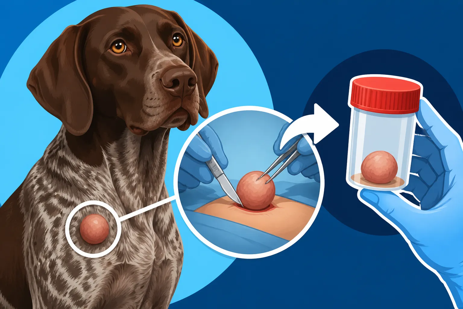
There are two main types of surgical biopsies: an incisional biopsy, where only a piece of the tumor is removed, and an excisional biopsy, where the entire lump is removed with a margin of surrounding healthy tissue. The choice depends on the size, location, and suspected nature of the mass. An excisional biopsy is often preferred if the vet suspects malignancy, as it serves both diagnostic and therapeutic purposes, potentially removing all cancerous cells in one go. After the surgery, your German Shorthaired Pointer will need proper care, including pain management and preventing them from licking or chewing at the incision site (which might mean wearing an "e-collar" or a soft recovery suit, easily found on sites like Chewy). The tissue is then carefully preserved and sent off to a specialized laboratory for a definitive diagnosis.
Understanding the Pathology Report and Future Care
Once your veterinarian has collected the necessary tissue sample—whether through an FNA or a surgical biopsy—the critical next phase begins: pathology. This is where the true nature of your German Shorthaired Pointer's lump will finally be revealed. Waiting for these results can be agonizing, a period of heightened anxiety, but understanding what happens during this stage and how to interpret the report will be incredibly helpful.
The Journey of the Tissue Sample to the Lab
After your vet has performed the biopsy or FNA, the collected tissue is carefully prepared. For surgical biopsies, the tissue is typically placed in a special preservative solution, like formalin, which stops cellular decomposition and preserves its structure. FNA slides are air-dried or fixed. These samples are then packaged securely and sent to a veterinary diagnostic laboratory. These labs are staffed by highly trained veterinary pathologists who specialize in examining animal tissues and cells for disease. They meticulously analyze the sample under powerful microscopes, looking for abnormalities in cell structure, arrangement, and staining characteristics. They are the detectives of the microscopic world, piecing together clues to give a definitive diagnosis. This is an essential step, as it removes much of the guesswork and provides a clear path forward.
For example, a pathologist might look for the characteristic round cells of a mast cell tumor, or the spindle-shaped cells of a fibrosarcoma. They can identify benign growths like lipomas (which are simply collections of fat cells) or sebaceous cysts (blocked oil glands), distinguishing them from aggressive cancers. This detailed examination is far more comprehensive than what can typically be done in a general veterinary clinic, making the specialized lab a crucial partner in your GSP's care.
Decoding the Results and Planning Your Shorthair's Next Steps
When the pathology report arrives back at your vet's office, it will contain a wealth of information. Your vet will sit down with you to explain the findings in detail, translating the medical jargon into understandable terms. The report will definitively state whether the lump is benign (non-cancerous) or malignant (cancerous). If it's malignant, it will often specify the type of cancer, its grade (how aggressive it appears), and whether the surgical margins were clear (meaning all cancerous cells were removed with a border of healthy tissue).
For instance, if the report identifies a Mast Cell Tumors and Where They Appear on a Shorthair, it might also indicate the grade of the tumor, which guides the prognosis and further treatment. If it's a low-grade tumor with clear margins, the removal might be curative. If it’s high-grade or has incomplete margins, additional treatments like radiation, chemotherapy, or further surgery might be recommended. This is where the decisions about your GSP's future care become very real. Your vet will discuss all options, potential outcomes, and what these mean for your dog's quality of life. This could involve anything from monitoring for recurrence to consulting with a veterinary oncologist for advanced cancer treatment plans. For more on surgical options, see Removing a Mast Cell Tumor From a German Shorthaired Pointer. Even if the diagnosis is tough, having a clear understanding of the situation empowers you to make the best, most informed choices for your loyal companion.
Managing the Costs of Lump Removal in Your German Shorthaired Pointer
Let's be honest, getting a diagnosis and treatment for a lump on your German Shorthaired Pointer, especially if it requires surgery and specialized lab work, can come with a significant financial cost. This isn't just about the emotional toll; the practical reality of veterinary bills can be a huge source of stress for many pet owners. It’s important to understand the various components that contribute to the overall expense so you can prepare as best as possible.
Anticipating the Financial Impact of Diagnosis and Surgery
The cost for addressing a lump on your GSP can vary widely based on several factors, including your geographic location, the specific veterinary clinic, the size and location of the lump, the complexity of the surgery, and the type of diagnostic tests performed. An initial vet visit and a fine needle aspirate are generally the least expensive steps. However, if a surgical biopsy or complete removal is needed, the costs will naturally escalate.
Here's a breakdown of common cost components:
- Initial Vet Exam: For the palpation and initial assessment.
- Fine Needle Aspirate (FNA): Includes the procedure itself and the pathology reading.
- Pre-surgical Bloodwork: Essential to ensure your German Shorthaired Pointer is healthy enough for anesthesia. This also helps assess organ function.
- Anesthesia: This is a significant part of any surgical procedure, involving medications, monitoring, and the time of the veterinary staff. Anesthesia Risk in a Lean German Shorthaired Pointer is a good read.
- Surgical Removal: The actual cost of the procedure, which varies based on the size, depth, and complexity of the lump. Lumps in difficult areas might require more specialized surgery.
- Post-operative Medications: Pain relief, antibiotics, and sometimes anti-inflammatory drugs.
- Pathology Report: The cost for the specialized lab to analyze the tissue and provide a definitive diagnosis.
- Follow-up Visits: To check the incision site and discuss results.
It's crucial to get a detailed estimate from your vet before proceeding with any surgery. Don't hesitate to ask questions about every line item so you understand what you're paying for. While we can't give specific figures here, rest assured that lump removal can range from a few hundred dollars for a simple benign removal to several thousand for a complex, cancerous tumor requiring extensive margins and specialized care. For a broader look at vet expenses, consider The Vet Bills a Hunting German Shorthaired Pointer Earns.
How Pet Insurance Can Ease the Burden of Unexpected Vet Bills
This is precisely where pet insurance can be a lifesaver. Many owners don't consider pet insurance until an emergency strikes, but having a policy in place before your German Shorthaired Pointer develops a lump can make a monumental difference in your financial stress and your ability to choose the best treatment options. Providers like Healthy Paws offer comprehensive accident and illness plans that typically cover diagnostics, surgery, medications, and even specialist care related to new medical conditions, including lump removal and cancer treatment.
If your GSP has a pre-existing condition, it might not be covered, which is why early enrollment is so beneficial. Always review policy details carefully to understand deductibles, reimbursement percentages, and annual limits. Even if you don't have insurance, it's wise to have a financial plan for unexpected veterinary expenses. This could mean setting aside an emergency fund, investigating veterinary credit lines (such as The Credit Line a Shorthair Owner Should Arrange), or discussing payment plans with your vet. While some people consider self-insuring, unforeseen, high-cost events like cancer treatment can quickly deplete savings that might not have grown enough. Having that safety net can allow you to focus on your dog's recovery without the added worry of a crushing financial blow. For more on general insurance considerations, look at Monthly Insurance Premiums on a German Shorthaired Pointer.
Comparing Diagnostic Approaches for German Shorthaired Pointer Lumps
Here's a quick overview of the two primary diagnostic methods discussed, to help clarify their differences and when each might be used:
| Feature | Fine Needle Aspirate (FNA) | Surgical Biopsy |
|---|---|---|
| Invasiveness | Minimally invasive | Moderately invasive |
| Anesthesia | Usually none or mild sedation | Required general anesthesia |
| Cost | Generally lower | Generally higher (includes surgery, anesthesia, etc.) |
| Information Provided | Cellular-level diagnosis, can differentiate fat, inflammation, mast cells. | Definitive tissue diagnosis (histopathology), tumor type, grade, margin assessment. |
| Recovery Time | Immediate | Days to weeks, requires wound care |
| Limitations | May yield insufficient or non-diagnostic samples, can miss certain tumor types. | More expensive, requires anesthesia, longer recovery. |
| When Typically Used | First line for palpable lumps, screening for benign conditions. | When FNA is inconclusive, or high suspicion of malignancy, or for complete removal. |
It's important to remember that these tools are often used in conjunction, with an FNA providing initial guidance and a surgical biopsy offering the definitive answer when needed. Regular hands-on checks of your German Shorthaired Pointer, perhaps after a hunt as outlined in Checking a German Shorthaired Pointer Over After a Hunt, can help you find these lumps early when they are smaller and potentially easier to manage.
Disclaimer
This article is for general informational purposes only and does not constitute professional advice. Laws, prices and regulations vary by location and change over time. Consult a qualified professional for guidance on your specific situation.